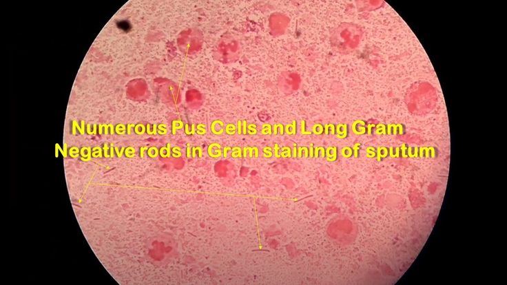
Capsule Staining India Ink
Demonstration can be facilitated by staining them with india ink injected through the genital pores. Web Study with Quizlet and memorize flashcards containing terms like 1.

Tinciones Para Bacterias Medical Laboratory Science Medical Laboratory Microbiology
The ability of fungi to invade plant and animal tissue was observed in early 19th century but the first documented animal infection by any fungus was made by Bassi who in 1835 studied the muscardine disease.

. All e-mails from the system will be sent to this address. Web We would like to show you a description here but the site wont allow us. This is not a true staining method but highlights the capsule as a transparent halo around the bacillus.
Web Advanced search for Ongoing Bids. Web Negative staining. No the saline mount would yield the same results as the iodine mount.
It is satisfactory with good capsule preparations such as blood from a freshly dead animal or smears of bacilli from mucoid colonies on bicarbonate agar grown under CO 2 see this annex section. Web We would like to show you a description here but the site wont allow us. Web India ink method for capsule visualization.
Web Provide a valid e-mail address. Web The cell envelope is composed of the cell membrane and the cell wallAs in other organisms the bacterial cell wall provides structural integrity to the cell. There is a dense milk white spot at the side where the scolex with its hooks and suckers remain invaginated.
Web Drugs Available In Health Facility Refresh Data Filter Data Download PDF. The background gets stained black whereas the unstained bacterial or yeast capsule stands out in contrast. Pylori hydrolyzes urea to ammonia which raises the pH of the medium and changes the color of the specimen from yellow.
Bid RA Number. This layer of polysaccharide sometimes proteins protects the bacterial cell and is often associated with pathogenic bacteria because it serves as a barrier against phagocytosis by white blood cells. Search by Ministry Organization.
After drying the microorganisms may be. A drop of bacterial suspension is mixed with dyes such as India ink or nigrosin. The test is performed at the time of gastroscopyA biopsy of mucosa is taken from the antrum of the stomach and is placed into a medium containing urea and an indicator such as phenol redThe urease produced by H.
The e-mail address is not made public and will only be used if you wish to receive a new password or wish to receive certain news or notifications by e-mail. When using an iodine mount to examine a fecal specimen for intestinal protozoa or helminth ova is it still necessary to perform a saline mount. In prokaryotes the primary function of the cell wall is to protect the cell from internal turgor pressure caused by the much higher concentrations of proteins and other molecules inside the cell compared to.
7-12 lateral branches on. Cryptococcus neoformans appears as spherical budding yeast forms 2-10 μ in diameter and surrounded by a large unstained capsule Figure 11828. Alternatively positive and negative staining techniques can be combined to visualize capsules.
Therefore mycology is the study of fungi. Web The latest Lifestyle Daily Life news tips opinion and advice from The Sydney Morning Herald covering life and relationships beauty fashion health wellbeing. Web In the spring of 2020 we the members of the editorial board of the American Journal of Surgery committed to using our collective voices to publicly address and call for action against racism and social injustices in our society.
Web A simple staining method for bacteria that is usually successful even when the positive staining methods fail is to use a negative stainThis can be achieved by smearing the sample onto the slide and then applying nigrosin a black synthetic dye or India ink an aqueous suspension of carbon particles. No the iodine mount is superior to a saline mount. Web INTRODUCTION TO MYCOLOGY The term mycology is derived from Greek word mykes meaning mushroom.
Other capsular stains can also be used to negatively stain encapsulated cells Figure 237. Capsules can be seen by viewing bacteria in India ink. Web The cyst is separated from the host tissue by a thin collagenous capsule.
Web Alternatively a drop of India ink is added to the drop of sediment on a glass slide a coverslip is placed and examined under the microscope using 40 objective. Web One common negative staining technique for identifying encapsulated yeast and bacteria is to add a few drops of India ink or nigrosin to a specimen. This is very useful in the demonstration of capsules that do not take up simple stains.
Search by Bid RA Details.
Pin On Bacteriology

Pin By Mayra Rolon On Medical Microbiology Medical Laboratory Science Microbiology Medical Laboratory

Bacillus Megaterium Capsule Stain With India Ink And Crystal Violet India Ink Ink Crystals
Rosh Review India Ink Stain Microbiology Infectious Disease
0 Response to "Capsule Staining India Ink"
Post a Comment